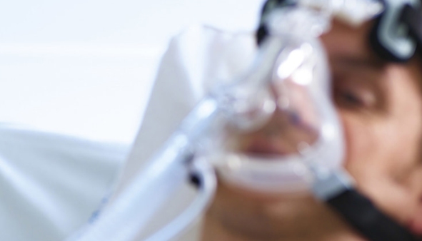
Children's airways are narrower and more flexible compared to adults, so they are more susceptible to obstruction and respiratory issues. When such problems arise, pediatric airway management techniques can prevent sudden death. In most cases, medical staff can help maintain airway patency through routine methods like airway clearance and respiratory support. Therefore, it is important to select the right airway management ways. This article will introduce common airway management methods, let's have a look at the key features of pediatric respiratory function first. Key Physiological Features of Pediatric Respiratory Function Lung Capacity: Children have lower lung volumes compared to adults, which gradually increase with age. Tidal Volume: Younger children always have smaller tidal volumes. Airway Resistance: Children's airways have higher resistance, especially in cases of small airways, which makes airway obstruction more likely. Respiratory Reserve: Children have lower respiratory reserve and their breathing function is relatively fragile. Minute Ventilation: The minute ventilation (calculated by body surface area) is similar in adults but overall lower in children. Functional Residual Capacity: Children's lungs have less reserve capacity than adults. Gas Diffusion Capacity: The total surface area of capillaries in a child's lungs is smaller, which results in a lower gas diffusion capacity. Lung Elasticity: Children’s lungs have less elasticity, which limits their lung volume and recovery. Incomplete Alveolar Development: Newborns have only 8% of the alveoli found in adults, and their lungs are still developing, leading to less respiratory reserve. Anatomical and Physiological Features of Pediatric Airways Children's heads are disproportionately large, with shorter necks, larger tongues, and more prominent occiputs, which can cause airway constriction. Newborns and infants up to 5 months of age predominantly breathe through their noses, making nasal patency critical. Enlarged tonsils and adenoids in preschool children can cause upper airway narrowing. Generally, the epiglottis in children is epiglottis-shaped, which can affect intubation techniques. Newborns have limited alveolar numbers and less lung development, reducing their capacity for reserve breathing. Common Airway Management Methods 1. Face Mask A face mask is the most basic method of ventilation and is widely used in pediatric anesthesia. However, in newborns and infants under 5 months, it can be challenging due to its potential to compress the nasal passages. Additionally, in preschool children with enlarged tonsils or adenoids, face mask ventilation may be more difficult. Prolonged mask use can also increase the risk of gastric insufflation and aspiration. 2. Oropharyngeal Airway This airway is used to relieve upper airway obstruction due to an oversized tongue, particularly during anesthesia. It is crucial to select the correct size when using an oropharyngeal airway. By measuring the distance from the corner of the mouth to the angle of the jaw or earlobe, you can get the right size. Importantly, it must be placed at an appropriate depth to avoid obstructing the airway or pushing the epiglottis over the glottis, which could affect ventilation. It's also to be cautious as it can cause injury to the tongue or teeth, or slip out of position. 3. Nasopharyngeal Airway NPA can relieve upper airway obstruction and maintain airway patency. However, this method can damage the mucosa in the nasal passages, especially in children with preexisting nasal ulcers or necrosis. The device may also accidentally slip into the esophagus, leading to inadequate ventilation and gastric insufflation. 4. Endotracheal Intubation Endotracheal intubation is the standard for ensuring ventilation during general anesthesia. It provides an excellent oxygen supply during surgery and reduces the risk of aspiration. However, the anatomical features of children such as larger heads, shorter necks, and higher larynx position make intubation challenging. The procedure can cause trauma to the soft tissues of the larynx and lead to edema, resulting in respiratory distress. Moreover, intubation can have great effects on hemodynamics, causing increased blood pressure and heart rate, it may disrupt the normal course of surgery. 5. Laryngeal Mask Airway (LMA) The LMA combines the advantages of both face masks and endotracheal tubes and is widely used in pediatric anesthesia. Compared to traditional intubation, LMA insertion is easier and doesn't require a laryngoscope. This method is also faster, and it creates a secure airway without causing trauma to the larynx and trachea. Studies show that LMA anesthesia is more effective in young children undergoing day surgery, as it better maintains hemodynamic stability and reduces the occurrence of respiratory complications and hypoxia. Conclusion The anatomical and physiological features of children's airways make airway management complex. Therefore, it is crucial to select the right method of airway management, helping quickly and effectively resolve airway issues and ensuring the child's respiratory safety. When choosing an airway management product, it's recommended to use reliable and designed to suit the anatomical features of children. Bever Medical provides ideal airway management products to better accommodate children's anatomical structures, improving safety and ease of use. Any interests, welcome to contact us.
View More +-
10 Dec 2024
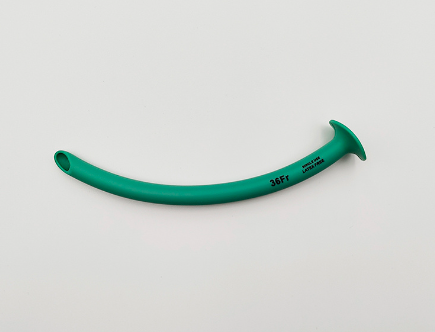
There are many materials for nasopharyngeal airways, PVC and silicone are two of the more common choices. Due to its unique performance characteristics, PVC nasopharyngeal airways have obvious advantages but also have disadvantages in patient use. Today, Bever Medical will share with you the benefits and limitations of PVC materials, as well as how to maintain them. Nasopharyngeal Airway (NPA) A nasopharyngeal airway, also known as a nasopharyngeal tube, is a hollow, soft tube with a smooth interior, designed for simplicity and ease of use. A PVC nasopharyngeal airway refers to one made of polyvinyl chloride (PVC), categorized as a non-endotracheal airway device. Typically, it is placed outside the glottis, creating a passage in the nasopharynx. This tube helps support collapsed soft tissues, advances the tongue base forward to relieve airway obstruction, clear secretions, and maintain airway patency. Compared to oropharyngeal airways, nasopharyngeal tubes cause less irritation to the throat, offering greater comfort for patients during use. Structure of the Nasopharyngeal Airway The nasopharyngeal airway consists of three main components: Connector: Features a cavity running through its length, allowing for the insertion of a catheter to pass through the airway. The upper end has a conical interface to connect and secure oxygen supply tubing, while the lower end fits tightly with the sleeve. Sleeve: A hollow tube that connects and fixes the tube and the connector, securing it to the tube’s opening. Tube: One end of the tube is beveled for easier insertion, while the other end is fitted with a sleeve for stabilization. Benefits of PVC Nasopharyngeal Airways 1. Good Biocompatibility and Durability PVC's biocompatibility makes it suitable for short-term use, as it generally does not cause significant tissue damage. This quality contributes to its widespread application in medical devices. Its durability also makes it ideal for various environments, including emergency care and short-term medical interventions. However, long-term use might increase the risk of infection or tissue damage. 2. Ease of Use The moderate rigidity of PVC nasopharyngeal airways, combined with their simple design, makes them easy to insert and operate, especially for emergency medical personnel and beginners. It is crucial to use appropriate lubrication and follow proper techniques to minimize patient discomfort and potential irritation. 3. Low Cost The low production cost of PVC makes it a preferred material for disposable medical devices, offering a significant price advantage for B2B procurement needs. Compared to higher-cost materials like silicone, PVC is more suitable for short-term use. 4. Adaptability to Various Environments PVC nasopharyngeal airways maintain stable performance in challenging environments such as high temperatures and humidity. This makes them particularly useful for emergency medical situations and field rescue operations. However, extreme temperatures may slightly affect the material’s flexibility. For specific needs, you can consult suppliers like Bever Medical, which provides high-quality nasopharyngeal airways for medical and field rescue applications. Limitations of PVC Nasopharyngeal Airways 1. Potential Discomfort or Allergic Reactions Although PVC has good biocompatibility, additives such as plasticizers may trigger allergic reactions in some patients, especially those with hypersensitivity. Physicians should evaluate the patient’s medical history for allergies and consider alternative materials like silicone or latex for sensitive individuals. 2. Risk of Infection or Airway Damage from Prolonged Use The rigidity of PVC compared to silicone may cause mechanical damage to nasal and airway tissues during long-term use. Additionally, prolonged placement increases the risk of bacterial colonization, leading to infection. Therefore, PVC nasopharyngeal airways are best suited for short-term applications, with strict adherence to aseptic protocols. 3. Lower Comfort Compared to Other Materials PVC nasopharyngeal airways are less flexible and comfortable than silicone or other materials. For patients requiring long-term use or prioritizing comfort, silicone options may be more appropriate. Specific Patient Groups for PVC Nasopharyngeal Airways Patients with Locked Jaw: For patients unable to clear secretions through the mouth, a nasopharyngeal airway can help maintain airway patency. Patients with Pharyngeal Tumors: Nasopharyngeal airways are beneficial for those with oropharyngeal tumors causing swallowing difficulties or airway obstruction. Patients with Ineffective Coughing and Secretion Clearance: Suitable for cases such as acute exacerbation of chronic obstructive pulmonary disease (AECOPD) or post-extubation situations to prevent repetitive nasal mucosal damage during suctioning. PVC Nasopharyngeal Airway (Trumpet Type) Maintenance and Cleaning Requirements for PVC Nasopharyngeal Airways 1. Disposable vs. Reusable Products Disposable products must be discarded immediately after use to avoid cross-infection. For reusable products, rinse off residue before cleaning. Sterilization methods should align with the material, such as high-temperature steam sterilization or chemical disinfectants like phenolic compounds or hypochlorite solutions. Ensure complete sterilization before reuse. 2. Aseptic Conditions Insert and use the device under sterile conditions to prevent contamination. Inspect the airway for damage, blockages, or expiration before use. Replace any defective products promptly. Conclusion PVC nasopharyngeal airways are widely used in the medical field due to their low price and easy operation, but they also have limitations. For example, long-term use may cause discomfort or infection, so it is very important to choose the right material. For patients, the right choice of nasopharyngeal airways can effectively keep the airway open, relieve symptoms, and help treatment. If you don't know which is best for you, it is a wise way to consult your doctor, he or she will recommend it according to your situation. Additionally, if it is difficult for you to select the size of the NPA, you can ask suppliers such as Bever Medical for more advice on medical materials and products. We provide nasopharyngeal airways in a variety of materials and can provide you with detailed instructions for use to ensure that you make the best choice.
View More + -
04 Dec 2024
Airway management is a cornerstone of emergency medicine, anesthesia, and critical care. Nasopharyngeal airways (NPA) and oropharyngeal airways (OPA) are two common tools used to secure the airway, ensuring adequate ventilation and oxygenation. While both devices serve the primary purpose of maintaining a patent airway, they differ significantly in design, use cases, and clinical application. This document explores these differences in detail to help clinicians choose the most appropriate tool for a given situation. 1. What is an NPA? An NPA, or Nasopharyngeal Airway, is a flexible, tubular device inserted through the nostril into the nasopharynx to maintain airway patency. It is designed to bypass obstructions in the oropharynx and create a channel for airflow. Key Features of NPA: Material: Usually made of soft, pliable rubber or silicone to reduce trauma during insertion. Shape and Design: Cylindrical with a flared end to prevent complete insertion and a beveled tip for easier navigation through the nasal passages. Sizes: Available in various lengths and diameters to suit different patient anatomies, from pediatric to adult. Applications: Suitable for patients with an intact gag reflex or those who are semi-conscious. Commonly used in prehospital care, anesthesia, and intensive care units. Indicated for situations where oral airway insertion is contraindicated, such as facial trauma, clenched jaw, or limited mouth opening. BEVER Medical Nasopharyngeal Airway (NPA) is a high-quality, medical-grade device designed to provide secure and comfortable airway management. Crafted from medical-grade PVC material, this NPA is specifically engineered to meet the needs of emergency medical professionals, paramedics, and healthcare providers. Its thoughtful design, coupled with advanced materials, ensures both safety and comfort for patients in critical care situations. 2. What is an OPA? An OPA, or Oropharyngeal Airway, is a rigid device inserted through the mouth to prevent the tongue from obstructing the oropharynx. It is primarily used in unconscious patients who lack a gag reflex. Key Features of OPA: Material: Rigid plastic or rubber. Shape and Design: Curved to follow the anatomy of the palate and pharynx, with a flange to prevent over-insertion. Sizes: Available in various lengths to accommodate pediatric and adult patients. Applications: Commonly used in emergency settings, anesthesia, and airway management during cardiopulmonary resuscitation (CPR). Indicated for unconscious patients with no gag reflex to prevent airway obstruction caused by the tongue or soft tissues of the mouth. 3. Key Differences Between NPA and OPA 4. Advantages and Disadvantages NPA Advantages: Can be used in patients with intact gag reflex. Effective for conscious or semi-conscious individuals. Less risk of inducing vomiting or laryngospasm compared to an OPA. NPA Disadvantages: Risk of nasal trauma or bleeding during insertion. Contraindicated in basal skull fractures due to potential brain injury. May cause discomfort or irritation, especially in long-term use. OPA Advantages: Simple and quick to insert with minimal training. Highly effective in maintaining airway patency in unconscious patients. Prevents tongue-related airway obstruction during resuscitation or surgery. OPA Disadvantages: Cannot be used in conscious patients or those with an intact gag reflex. Risk of inducing vomiting or laryngospasm if used incorrectly. Not suitable for patients with oral trauma or jaw clenching. 5. Choosing Between NPA and OPA The choice between NPA and OPA depends on the patient's condition, level of consciousness, and the clinical scenario: Use NPA when: The patient is conscious or semi-conscious. There is a need to maintain airway patency in the presence of oral trauma or trismus (jaw clenching). The patient has a gag reflex that contraindicates OPA use. Use OPA when: The patient is unconscious and lacks a gag reflex. The primary cause of airway obstruction is the tongue or soft palate. A simple, quick solution is needed during resuscitation. 6. Conclusion NPA and OPA are both essential tools in airway management, each with specific indications and contraindications. Understanding their differences, appropriate use cases, and insertion techniques is crucial for effective airway management. Proper assessment of the patient's condition and anatomy will guide clinicians in selecting the most suitable device, ensuring optimal outcomes in emergencies and critical care scenarios.
View More + -
27 Nov 2024
In order to make safer and more effective decisions quickly in emergency situations, it is very necessary to know the applicability of the two airway tools. This is very helpful for first responders, medical staff or ordinary people who want first aid knowledge. As a supplier of airway management products, Bever Medical will share with you when to use NPA instead of OPA today. Differences Between NPA and OPA Functions Nasal pharyngeal airways (NPA) and oropharyngeal airways (OPA) are commonly used airway management products in clinical practice to maintain airway patency. They share similar purpose, but they differ significantly in design, mechanism of action, and applicable scenarios: 1. NPA Functions NPA (Nasal pharyngeal airways) is a soft, tubular device inserted through the nasal passage to extend into the nasopharynx, creating an artificial airway. It bypasses soft tissue obstructions in the upper airway, ensuring airflow in cases where the patient cannot open their mouth. Its flexible material and lubricated surface enhance patient comfort during insertion, making it particularly suitable for patients with intact gag reflexes. 2. OPA Functions OPA (oropharyngeal airways), made of rigid, curved plastic, is inserted through the mouth and positioned along the tongue to reach the posterior pharyngeal wall. It prevents airway obstruction by displacing the tongue anteriorly, thus maintaining a clear passage. Due to its firm design and direct interaction with the tongue, OPA is most appropriate for unconscious patients without a gag reflex, as its use in conscious individuals may induce discomfort or vomiting. Pre-Lubricated Nasal Airway Scenarios for Using NPA 1. Patients Unable to Open Their Mouths NPA is ideal for patients with clenched jaws or oral injuries, such as those experiencing a seizure. It provides an alternative airway route through the nasal passage when the oral route is inaccessible. 2. Patients with an Intact Gag Reflex For conscious or semi-conscious patients, OPA is often intolerable due to its rigid structure and potential to trigger the gag reflex. NPA, being softer and less invasive, is a more comfortable and viable option. 3. Presence of Facial or Oral Trauma In cases of facial burns, maxillofacial fractures, or oral trauma where the use of OPA is contraindicated, NPA can bypass the affected area and provide effective airway support. 4. Temporary Airway Support NPA is frequently used in emergency situations requiring short-term airway management, such as during CPR or at accident scenes, due to its quick insertion and ease of use. Scenarios for Using OPA 1. Unconscious Patients OPA is highly effective for unconscious patients, such as those under general anesthesia or in cardiac arrest, as it prevents the tongue from collapsing and obstructing the airway. 2. Patients Without a Gag Reflex Since OPA contacts the posterior pharyngeal wall, it may provoke gagging or vomiting in patients with an intact reflex. Therefore, it is only suitable for unconscious individuals lacking this reflex. 3. Long-Term Airway Support In scenarios requiring a stable airway for a prolonged period, such as intraoperative or postoperative care, OPA ensures a reliable airway while allowing easy access for healthcare monitoring. 4. Emergency Airway Management In hospital or pre-hospital settings, OPA is often the first choice for managing airway obstruction in unconscious patients due to its rapid deployment and effectiveness. Factors Influencing the Choice Between NPA and OPA Selecting between NPA and OPA requires a comprehensive assessment of the patient's condition. Key factors include: 1. Patient’s Level of Consciousness For conscious or semi-conscious patients, NPA is preferred as it avoids stimulation of the posterior pharyngeal wall, reducing discomfort. Conversely, OPA is better suited for unconscious patients requiring stable airway support. 2. Anatomical Considerations Structural anomalies, such as nasal septal deviation, may hinder NPA insertion. Similarly, oral abnormalities, such as maxillofacial trauma or severe dental malocclusion, may limit OPA use. 3. Urgency of the Situation In critical airway emergencies, such as cardiac arrest, OPA is often chosen for its rapid insertion and immediate resolution of obstructions. If the mouth cannot be accessed, NPA becomes the only viable option. 4. Patient History Patients with a history of nasal bleeding or nasal surgery may not be suitable for NPA. Similarly, individuals allergic to latex should avoid using latex-based OPA devices. Risks and Precautions for Using NPA and OPA 1. Risks Associated with NPA Nasal Bleeding: Improper insertion or fragile nasal mucosa can cause bleeding. Adequate lubrication and avoiding forceful insertion minimize this risk. Sinus Infection: Prolonged NPA use increases the risk of infection, necessitating regular replacement and sterile handling. Misplacement: Incorrect placement may result in airway obstruction or even aspiration. 2. Risks Associated with OPA Vomiting and Aspiration: OPA may induce gagging and vomiting in patients with a functional gag reflex, potentially leading to aspiration of gastric contents into the lungs. Oral Trauma: Excessive force during insertion may damage teeth or oral tissues. Worsened Obstruction: Incomplete insertion of OPA can push the tongue backward, exacerbating airway blockage. 3. Common Precautions Both two airway management products must use appropriately sized devices to ensure effective and comfortable insertion. Closely monitor the patient's breathing post-insertion to confirm airway patency. Adhere to strict aseptic techniques to prevent infections. Conclusion Although this article provides the situation of choosing NPA instead of OPA, there are some situations that need to be combined with patient needs. Simply put, NPA is more suitable for patients with trismus, gag reflex, or facial trauma. Additionally, high-quality airway management products like those provided by Bever Medical are made of safe PVC and silicon medical materials. And OE services can meet the needs of patients and provide them with suitable materials and designs, providing safer and more comfortable solutions for airway management.
View More + -
20 Nov 2024
Managing a nasopharyngeal airway (NPA) is a critical skill for healthcare professionals, it helps patients maintain an open airway. However, if performed incorrectly, it can endanger the patient’s life. As a trusted NPA manufacturer, Bever Medical knows the importance. Therefore, we share a comprehensive guide on how to manage a nasopharyngeal airway today. What Is a Nasopharyngeal Airway (NPA)? A nasopharyngeal airway (NPA) is used to relieve upper airway obstructions caused by tongue collapse. The traditional NPA resembles a tracheal tube. Its distal end has a short, rounded, beveled edge to reduce throat irritation, while the nasal end features a flange to prevent it from slipping into the nasal cavity. The traditional design of the PVC nasopharyngeal airway resembles a tracheal tube. Its distal end features a short, rounded, beveled edge to reduce throat irritation, while the nasal end includes a flange to prevent it from slipping into the nasal cavity. Compared to other devices, such as oropharyngeal airways, the PVC nasopharyngeal airway offers better patient tolerance and helps maintain more stable hemodynamic conditions, making it a reliable choice for airway management. Choosing the Right Size and Equipment The NPA's size is determined by its internal diameter and length, typically measured in millimeters. Choosing the right size is essential to ensure the airway remains open. Only a properly sized NPA can support the posterior pharyngeal wall, relieve obstructions, and maintain an open airway. The ideal insertion depth places the distal end within 3 cm of the epiglottis. While diameter is important, studies suggest that length has a greater impact on effectiveness. Therefore, select the size primarily based on the required depth of insertion. How to Manage a Nasopharyngeal Airway Managing an NPA involves selecting the right size, preparing the airway, and ensuring proper positioning. Below are detailed steps and precautions: Steps: 1. Choose the correct size NPA: Measure the distance from the tip of the nose to the earlobe to estimate the appropriate length. 2. Check the nasal passage: Ensure there are no obstructions, such as nasal polyps or a deviated septum. 3. Prepare the airway: Spray the nasal mucosa with a vasoconstrictor and local anesthetic (e.g., lidocaine or ephedrine solution). Clean the nostrils and lubricate the NPA’s exterior with paraffin oil or a suitable lubricant. 4. Insert the NPA: With the curved side facing the hard palate, gently guide the NPA along the nasal floor. Advance it until resistance is felt at the posterior pharyngeal wall. 5. Adjust as needed: Rotate the NPA 60–90 degrees to align it correctly and guide it into the pharynx. The airway should open, snoring should stop, and breathing should normalize. Tips Slightly tilt the patient’s head back during insertion for easier placement. When the NPA reaches the oropharynx, rotate it 180 degrees so the concave side faces downward, positioning the distal end behind the tongue. Precautions Avoid using NPAs on conscious or semi-conscious patients to minimize discomfort or risk of injury. Regularly check the NPA’s position, ensure it remains open and clean or replace it as necessary to prevent infections. Why Proper NPA Management Is Critical Effective management of an NPA is essential, especially in emergencies, due to the following benefits: 1. Restores airway patency quickly: NPAs can immediately relieve upper airway obstructions, preventing hypoxia. 2. Simple and versatile: NPAs are easy to use, widely applicable, and ideal for emergency and clinical settings. 3. Improves survival rates: Proper NPA use provides valuable time to save lives, especially in cases of tongue collapse or laryngeal obstruction. Common Challenges and Solutions Resistance Insertion may encounter resistance due to narrow nasal anatomy or insufficient lubrication. Solution: Check for obstructions like polyps or a deviated septum. Apply additional lubricant for smoother insertion. Bleeding The nasal mucosa may become irritated or injured during insertion. Solution: Use vasoconstrictors like diluted ephedrine to minimize bleeding risks. Avoid using oversized NPAs or applying excessive force. Discomfort or Gag Reflex Patients may experience discomfort or reflexive gagging. Solution: Apply a local anesthetic, such as lidocaine, to reduce sensitivity and insert the NPA gently to avoid overstimulating the patient. Ensuring Patient Safety and Comfort To ensure the procedure is safe and comfortable for the patient, follow these best practices: Choose the correct size: Measure carefully to select an NPA with the appropriate diameter and length. Prepare thoroughly: Clean the nasal passage and use sufficient lubrication and anesthetics. Handle gently: Avoid excessive force to reduce the risk of nasal mucosa injury. Monitor closely: Regularly check the patient’s reaction and adjust the NPA if necessary. Conclusion Choosing the right NPA and mastering its use is crucial for effective airway management. Bever Medical is dedicated to providing high-quality NPAs made from medical-grade materials. Our NPAs are scientifically designed to be flexible, durable, and safe, reducing operational risks and enhancing patient comfort. Whether for emergency care or routine clinical use, Bever Medical NPAs are the reliable choice. Explore our catalog today and get top-tier airway management solutions!
View More + -
13 Nov 2024
A nasopharyngeal airway (NPA) is used in emergencies to help open the patient's airway, ensuring unobstructed breathing. It is particularly suitable for semi-conscious or drowsy patients at risk of airway obstruction. In emergency settings, NPAs also help healthcare professionals in ventilation procedures. However, an NPA that is too long can lead to complications or injury. Today, Bever Medical will share the potential complications caused by an NPA that is too long, and guide on selecting the right size for airway management products. What is the Complication of an NPA that is too Long? Airway Obstruction If the NPA extends too far, it may press against the epiglottis or the posterior pharyngeal wall, causing severe airway obstruction and worsening breathing difficulties. Additionally, foreign bodies or dried secretions can become lodged in the airway tube, leading to airway blockage. In semi-conscious patients, inserting an airway tube may trigger coughing or laryngospasm, potentially resulting in severe airway obstruction, especially if the airway's tip contacts the epiglottis or vocal cords. Nasal Bleeding If the NPA is too long, it may exert excessive friction and pressure on the nasal cavity, causing nasal bleeding. Patients with coagulation issues or those on anticoagulants are at higher risk for severe nasal bleeding. If bleeding originates from the anterior nasal vessels, applying pressure to the nasal area may help; if the posterior nasal vessels are bleeding, it may be necessary to leave the NPA in place, suction the pharynx, and ensure adequate ventilation. In cases where bleeding persists, intubation may be considered. Central Nervous System Injury In patients with basal skull fractures, an excessively long NPA could penetrate the nasal cavity and reach the cranial cavity, leading to central nervous system injury and serious complications. Pharyngeal Ulcers and Necrosis Prolonged placement of an NPA, especially one that is too rigid or too large, may lead to ulcers in the nasal or pharyngeal area. Hard or oversized NPAs can cause sustained pressure on the nasal and pharyngeal mucosa, resulting in tissue compression and ischemia, leading to ulcers. Long-term placement also raises the risk of infection; caregivers should monitor for signs of pressure ulcers or sinusitis for prompt management. Risk of Aspiration or Ingestion Using an NPA that is too long may increase the risk of aspiration into the pharynx or trachea, potentially leading to obstruction of the lower respiratory tract or even ingestion into the stomach, which can cause choking or gastrointestinal injury. Moreover, inserting the airway may trigger a gag reflex, raising intracranial pressure, which can complicate or create new injuries. Care providers should carefully monitor the patient's condition to reduce aspiration risks, quickly clearing any potential contaminants if the patient begins to vomit or experiences airway bleeding. How to Prevent Complications When Using an NPA Choose the Correct Airway Tube Size Selecting the appropriate size and softness of the NPA is crucial to preventing complications. Oversized tubes can unnecessarily press on the nasal and pharyngeal mucosa, leading to ulcers, bleeding, or other injuries; undersized tubes may not effectively maintain airway patency. Softer materials are preferable for extended use, as they reduce irritation to the nasal walls, lowering the risk of ulcers and infection. Alternate Between Left and Right Nasal Passages For patients requiring long-term NPA use, alternating between the left and right nostrils daily can reduce the risk of tissue damage and ulcers from prolonged pressure on one side, allowing both nasal passages to rest and recover. Regularly Humidify the Tube and Nasal Passage Regular humidification of the NPA and nasal cavity helps maintain mucosal moisture, preventing dryness and irritation. Because dry environments can cause the nasal and pharyngeal mucosa to crack, increasing the likelihood of ulcers and infection. So, using humidifiers, nasal sprays, or saline to alleviate discomfort in the nasal and pharyngeal area. How to Select the Proper Size for an NPA? Length: Measure the distance from the patient's nostril to the earlobe. This approximate length helps ensure the NPA's tip reaches the posterior pharyngeal wall without being too long or too short. Diameter: The airway tube's diameter should match the patient's nostril size to avoid being too tight or too loose. Generally, the diameter should be close to the width of the patient's little finger. Sizing for Children: Extra care should be taken when selecting an NPA size for children. Smaller-diameter tubes are typically recommended, and the length should be confirmed to avoid excessive length that could irritate. Adjustment and Testing: It is wise to apply a lubricant before insertion, as it can reduce friction and discomfort. Observe the patient's reaction during insertion to ensure the appropriate fit and maintain an open airway. Final Thoughts To ensure the safety and comfort of patients using an NPA, healthcare providers should follow the proper insertion steps, select the correct tube size, and closely monitor the patient's condition. These precautions help reduce complications and improve emergency care outcomes. Selecting high-quality airway management products, like those offered by Bever Medical, provides reliable support for both medical personnel and patients. For more information, please contact our professional team to learn how to choose the right equipment for your emergency needs.
View More +


